Blood Clot Diagnosis
Blood clot diagnosis involves suspecting the clot, and then proving that it is there. The most common location for a blood clot is in the leg veins. And the most useful tool to diagnose blood clots in the legs is ultrasound.
When to Suspect a Blood Clot
Usually, we will suspect a blood clot when a person has typical symptoms. A person who has a deep vein thrombosis in a leg vein might complain of leg swelling and pain. On the other hand, pulmonary embolism symptoms include chest pain and shortness of breath.
But, it is important to know that as many as 30% of people with clots will not have any symptoms. Sometimes we find clots incidentally, when imaging is done for other reasons. In other instances, we look for clots even if there are no symptoms. For instance in chronically immobile persons.
There are several tools we can use to help us decide if a clot is likely or not. The two we use the most are the Wells criteria and the Geneva score.
Blood Tests
The main blood test that helps with blood clot diagnosis is called d-dimer. Basically, when clot forms, d-dimer levels go up. So low d-dimer levels suggest there is no acute clot. On the other hand, elevated d-dimer levels may suggest clots. But, there are many other reasons for d-dimer to be high. So it is more useful to find that it is low than to know what to do with high levels.
Ultrasound for Blood Clot Diagnosis
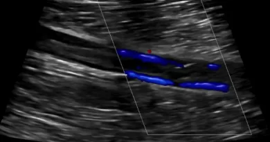
The main tool to diagnose a blood clot in a leg vein is ultrasound. The advantages of ultrasound are that it is accurate, safe, cheap and available almost everywhere.
There are three signs of clots that we can identify with ultrasound:
- Visible clot. Basically, this is when we can see the clot. But often we cannot actually see the clot. It is practically invisible on ultrasound.
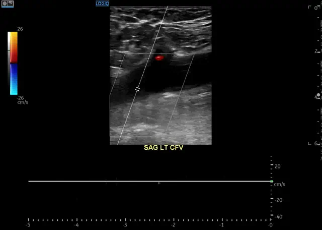
- Lack of blood flow. Veins are supposed to move blood up toward the heart. But if there is a blood clot in the vein, blood might not be able to flow freely. It is important to remember that some clots do not block flow completely. So even if we can detect blood flow, this does not mean there is nothing in the vein.
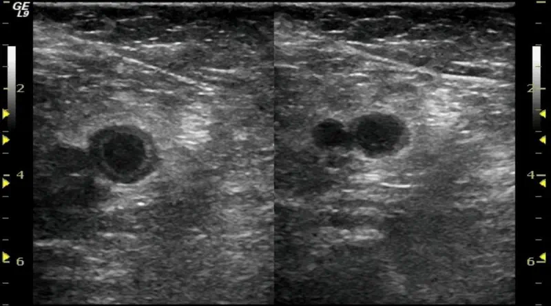
- Non-compressible veins. This is the most important finding. Veins are supposed to collapse when we press on them. But if there is clot in the vein, we will not be able to compress the vein. So even if we cannot see the clot, we will know it is there.
Blood Clot Diagnosis with CT or MRI
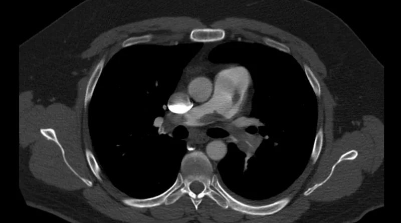
We usually diagnose blood clots in the lungs or in the abdominal veins with CT. Sometimes, we identify clots in the abdominal or pelvic veins with MRI. A special type of MRI called MRV is useful to diagnose iliac vein compression (May-Thurner Syndrome).
Echocardiogram
There are less common ways to diagnose clots. One is with an echocardiogram. While this study looks at the heart chambers and valves, it can also show clots in the heart. An echocardiogram is practically the most accurate way to diagnose a clot in transit. This is a clot on its way from the legs to the lungs, through the heart.
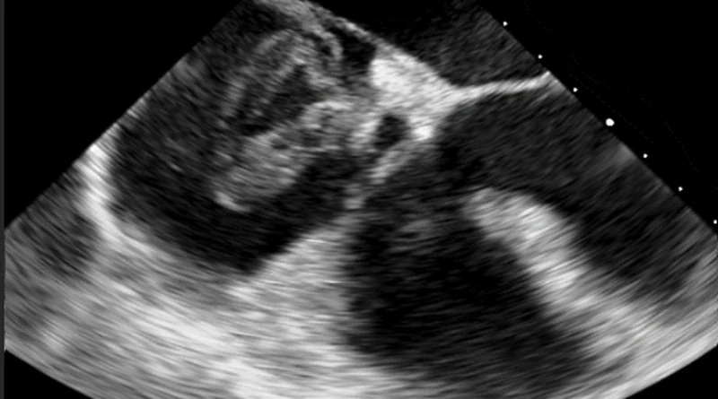
If we identify a clot in transit with echocardiogram, it is important to also look for a patent foramen ovale. A patent foramen ovale, or PFO, is a hole between the heart chambers. A clot in the heart may pass through a PFO. If that happens, it might skip the lungs and cause a stroke, which in the end is an artery blood clot.


Pingback: Is this Chronic Clot?
Pingback: Anticoagulation Failure: Definition and Causes