Is this Chronic Clot?
When we find a clot in a leg vein, knowing if it is fresh or chronic clot is not always easy. But it is still important to know. If the clot is fresh, we will usually need to treat the patient with blood thinners. But if the patient has chronic clot, then we may not have to. In real life, telling a fresh from a chronic clot is not always easy. Still, there are a few things to consider.
The Patient’s Story
Listening to patients is the most useful tool we have to diagnose disease. When a patient describes sudden onset of leg swelling and pain that they have never had before, we should think about a deep vein thrombosis. Then, if we scan their leg and we find evidence of a clot, we can assume it is acute (or fresh).
On the other hand, if a patient tells us that they had a clot in the past, chances are the clot we are looking at on the scan might be old.
But life is messier than these two scenarios. For instance, what if a patient had a clot in the past, but now we are suspecting a new clot? Another problem are post thrombotic syndrome symptoms. Patients who had a clot might develop long-term symptoms that might include leg swelling and pain. Of course, the symptoms of a deep vein thrombosis are also pain and leg swelling. Telling them apart can be challenging.
Blood Tests
A blood test called d-dimer is very useful when we are trying to understand how old a blood clot is. If the test shows a normal d-dimer, the chance of an acute clot is very low. But if the d-dimer is high, the clot might be fresh.
An important fact to understand about d-dimer, though, is that it can be elevated for many reasons. This means that d-dimer is great at ruling out a fresh clot. But if it is elevated, we should use other tools to make our decisions.
Ultrasound Characteristics of Chronic Clot
The test we use the most for blood clot diagnosis is ultrasound. In fact, ultrasound is very accurate in diagnosing deep vein thrombosis.

But on the other hand, ultrasound is not very accurate in determining clot age. Still, there are a few characteristics of clot that are more typical of chronic clot:
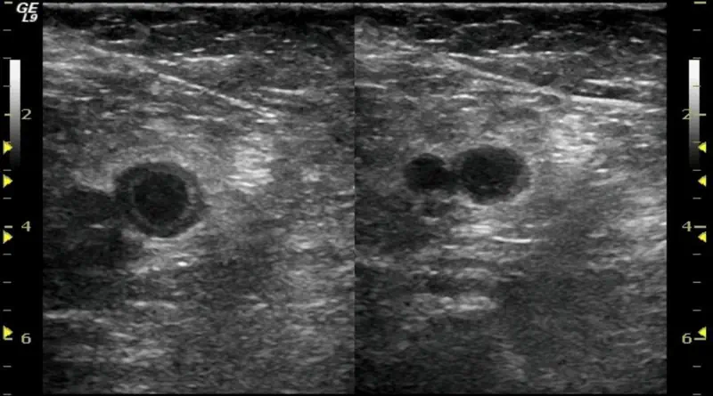
- Scarred vein. A scarred vein means the vein is shriveled and small. It takes veins time to get that way, so this will suggest a chronic clot.
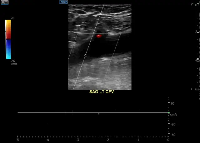
- Hyperechoic clot. This means you can see the clot easily with ultrasound. Typically, if the clot is “invisible”, then it is acute, or fresh.

- Recanalization. If there is some blood flowing through the vein, it might suggest the body had time to create new channels.
- Comparison to previous studies. If we have previous studies, we should always compare. Clot in the same place as an old study will support chronicicty.
The problem is, that when you show an ultrasound study to professionals, they don’t always agree on these characteristics.
Special Testing for Acute vs Chronic Clot
Because of the limitations of the patient’s story, blood tests and ultrasound, there is a need for better tools. There is much research looking into using special imaging studies to help determine clot age. The most obvious test is PET-CT. A PET-CT can “light-up” when there is inflammation. As an acute clot might cause inflammation of the vein, a PET-CT can show that. These techniques still have a long way to go and are not part of routine medical care.
Chronic Clot Pathology
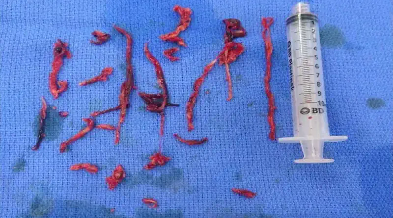
Sometimes we are able to obtain a sample of the clot. When this happens, we might be able to send the sample to pathology. Fresh clots are more red-blood cell rich. On the other hand, chronic clots are more fibrin rich. Even to the naked eye they will seem different. Fresh clots will look more red, while older clots will appear more white.

Pulmonary Embolism Clot Age
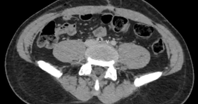
We should also mention pulmonary embolism. Similar to deep vein thrombosis, it is sometimes important to know if a pulmonary embolism is acute or chronic. Practically, we use similar tools as for deep vein thrombosis. First, we listen to the patient’s story. Sometimes, patients will describe typical symptoms, or tell us about an old clot that they had. Next, we might use d-dimer. As for DVT, a low d-dimer goes against an acute PE. Finally, we look at the CT scan. Similar to ultrasound, it is hard to determine clot age from a CT scan. But sometimes it is possible, especially by competent radiologists.


Pingback: Anticoagulation Failure: Definition and Causes